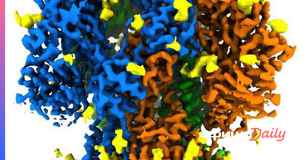
Scientists from the United States presented the first high-resolution images of the protein spines of the coronavirus. Images published on website National Accelerator Laboratory SLAC at Stanford University.
Coronaviruses have spikes that bind to receptors on the cells of their victims, leading to infection, the researchers said. Moreover, the virus is so active that there are only a few cryo-laboratories in the world that can study it with a sufficiently high level of safety for employees.
–
At the Stanford-SLAC Cryo-EM Facility, scientists studied a mild-mannered relative of the virus that causes #COVID19. The research paves the way for seeing more clearly how spike proteins initiate infections, with an eye to preventing and treating them. https://t.co/71uWKMmwNq
– SLAC (@SLAClab) December 18, 2020
–
–
“The advantage of our imaging is that when you purify the spine protein and study it yourself, you lose an important biological context: what does it look like in an intact virus particle? After all, there may be a completely different structure, ”- said Professor Wah Chiu of SLAC.
As part of the study, experts studied a milder strain of coronavirus called NL63, which causes the common symptoms of the common cold and causes about 10% of respiratory diseases every year. It attaches to the same receptors on the surface of human cells as COVID-19.
The experts did not chemically remove and purify NL63 proteins and instead froze the viruses whole to a glass state that preserves the natural arrangement of the components. They then took thousands of test images of randomly targeted viruses using cryo-EM tools and combined them to produce quality images.
Russian scientists done the first photo of the coronavirus in March. Specialists of the Research Center of Virology and Biotechnology “Vector” obtained images by negative contrasting on a JEM-1400 transmission electron microscope.
–
Preview image: SLAC
–
–


