Gent –
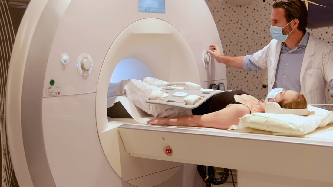
Radiologists at UZ Gent have succeeded in converting MRI images of bone structures into CT images in 3D with the help of advanced software. A breakthrough that can offer a solution to the dangerous X-rays that are released when taking classic CT images.
–
Every year, patients in Belgium undergo more than 2 million CT scans because precise imaging is necessary in the treatment of a bone injury. But the scans use X-rays, which makes …
–
Related posts:
COVID-19 Vaccination Program for Healthcare Workers and High-Risk Individuals: Boosters and Future P...
How a Spanish doctor carried out the first global vaccination campaign from 1803
Need for Quadrivalent Influenza Vaccines in Peru Highlighted by Civil Organization
Djokovic and Gignac, opposite reactions to the vaccine


