(wS / Uni) Siegen 05.10.2022 | Molecular biologist Prof. Dr. Hans Merzendorfer of the University of Siegen, together with an international team of scientists, gained new insights into the chitin formation process. The findings could contribute to better protection against insect-borne or fungal-related diseases.
Chitin is one of the most abundant biopolymers in nature. It is found, among other things, in the shells of insects and in the cell walls of fungi. Chitin is a renewable raw material with special properties that make it interesting, among other things, for medical applications. For this reason, scientists around the world are working intensively on the question of how chitin is formed and how it can be used for different purposes. An international team of molecular biologists with the participation of Siegen biology professor Dr. Hans Merzendorfer has now made a breakthrough in basic research: the scientists have used a method that allows them to observe the key enzyme for chitin production at the work. This gave them important insights into the chitin formation process. Their results were published in the current issue of the renowned scientific journal “Nature”.
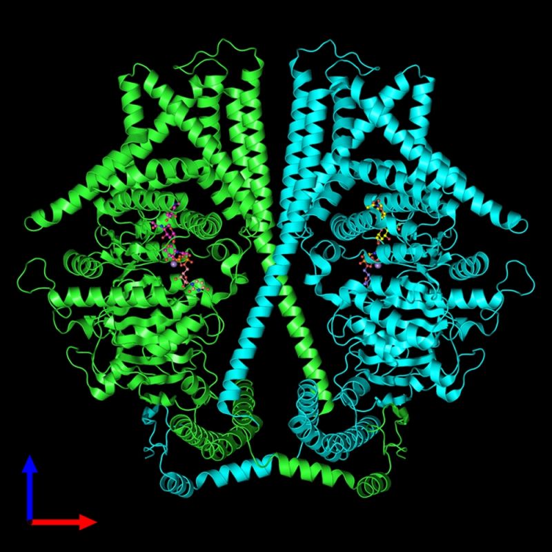
Chitin: 3D structural model of the key enzyme for chitin formation. The enzyme is present as a tightly intertwined complex of two molecules, each with a short chitin chain attached to the channel entrance

Merzendorfer: Molecular biologist Prof. Dr. Hans Merzendorfer of the University of Siegen, together with international colleagues, managed to observe the key enzyme for chitin production at work. Photo at the University of Siegen
“I am very happy with this publication, but especially with the new results of our research, which could help to better protect people from certain diseases in the future,” says Prof. Merzendorfer. We are talking about diseases such as malaria or yellow fever, which are transmitted by mosquitoes, but also about systemic fungal diseases that affect human organs and can be fatal. The fact that insects and fungi produce chitin in equal measure could be the decisive lever for combating these diseases: if the formation of chitin can be prevented in a targeted way, the massive spread of dangerous insects and fungi could be prevented.
“Even today, some insecticides or fungicides work by inhibiting the formation of chitin. However, this approach can be greatly improved through our knowledge of the structure and mode of functioning of the key enzyme “chitin synthase”: the better we understand the chitin biosynthesis process by this enzyme, the more specifically we can paralyze it by blocking the activity of chitin. ‘enzyme. “, explains Prof. Merzendorfer. He hopes that, on this basis, new and effective drugs against fungal diseases in humans can be developed in the future.” Since humans do not produce chitin on their own, we assume that the drugs that aim to block chitin biosynthesis should have fewer side effects, “explains the molecular biologist.
As part of their studies, the scientists took a close look at a special soybean rot fungus in the truest sense of the word. Using a high-resolution electron microscope that works in extremely cold conditions, they were able to visualize the atomic structure of chitin synthase at different times during the chitin formation process. To do this, the team first examined hundreds of thousands of individual particles with different spatial orientations. The bioinformaticians then used these images to compute a 3D model of the enzyme with atomic resolution.
“In this way, we were able to visualize various areas of the enzyme, including the catalytic center, by first connecting the individual sugar molecules to form a chain. The channel through which the growing chitin chain is transported was also visible. through the cell membrane to the outside. And we discovered a lock at the entrance to the canal, which also helps guide the chain into the canal, “says Merzendorfer. Another discovery: two key enzyme molecules are always involved in the formation of chitin. These are tightly entangled and produce two chitin chains in parallel, which should facilitate the formation of so-called chitin fibrils, i.e. bundles of different chitin chains.
In the next step, the team wants to work to broaden the knowledge gained and find out how they can be used for the production of improved chitin synthase inhibitors. In the future, such inhibitors could be used not only in medicine but also in agriculture to protect crops from insect or fungal infestations.
Nature’s current publication on the subject is available here: https://doi.org/10.1038/s41586-022-05244-5
.
announcement


