Since the new coronavirus was detected, worldwide research has been trying to find a way to control the virus. So far there are already several studies and even some interesting discoveries.
Through microscopic images it is possible to see the new coronavirus infecting cells.
Images were enlarged up to 200x to see the action of the coronavirus
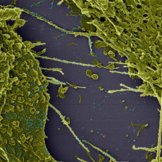
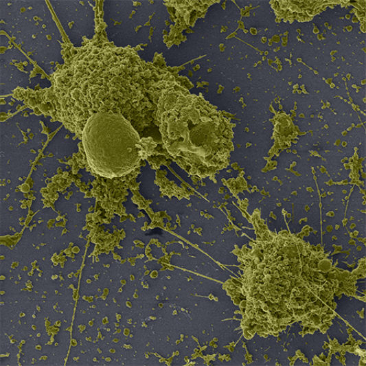
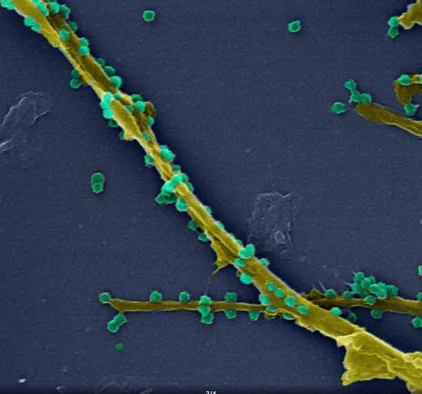
Using a high-resolution microscope, which can enlarge images up to 200,000 times, it is possible to have an impressive view of the way the virus behaves. The new images show, in great detail, how the new coronavirus, responsible for the disease COVID-19, infects cells.
In addition, it is also possible to see how viral particles are transmitted from one cell to another. The work is from the Oswaldo Cruz Foundation (Fiocruz) and the National Institute of Metrology, Quality and Technology (Inmetro), in Brazil.
According to the work done, it is possible to prove that the infected cells, in green, have membrane extensions, even 48 hours after infection. The researchers believe it is these extensions that allow the transfer of viral particles to adjacent cells.
Scientists say that this whole process is not very common, but it is similar to what happens in other infections, namely with the Ebola or Marburg virus.
The study was carried out with Vero cells, derived from monkey kidney and widely used in virology research. The images are preliminary results of the research project 'Study of the morphology, morphogenesis and pathogenesis of Sars-CoV-2 in in vitro, in vivo systems', carried out in collaboration between the Laboratory of Morphology and Viral Morphogenesis and the Laboratory of Respiratory Viruses and IOC measles.
The photographs were recorded with a high-resolution triple-ion microscope, in cooperation with researchers Braulio Archanjo and Willian Silva, from Inmetro's Electron Microscopy Laboratory.
-


