Created: 16.11.2024 | 08:30
Updated: 11/16/2024 | 09:26
Bacteriology marked the birth of a science. It arrived before the birth of virology, due to a matter of size. However, the study of viruses already has a long history, especially those viruses capable of causing diseases in humans. Although it is true that direct observation of the behavior of viruses has not always been an easy path, since most of them are too small to be seen with conventional optical microscopes, which are the first type of microscopes that were designed. This obstacle has limited our understanding of how viruses interact with their host cells and spread. The appearance of Mimivirusknown as the “giant virus”, has changed the landscape. This type of virus, discovered in 2003, has an unusually large size, allowing it to be seen through optical microscopes. He Mimivirus is he Diplodocus from the viral world.
In a recent study led by Kanako Morioka, Ayumi Fujieda y Masaharu Takemurait has been possible to film for the first time the infection process of the Mimivirus in cells of Acanthamoebaits natural host. They have used an innovative technique to record an educational sequence that shows the life cycle of the Mimivirus in real time. This film has not only attracted attention as a powerful visual tool for teaching biology, but also offers new insights into the interaction between giant viruses and their hosts.
He Mimivirus: A giant among viruses
He Mimivirusbelonging to the Mimiviridae family, is one of the largest viruses ever discovered. With a diameter ranging between 450 and 800 nanometersis much larger than most known viruses, allowing it to be observed with a standard light microscope. Its discovery revolutionized virology, as it challenged the classical definition of viruses, which were believed to be all too small to be visible with this type of microscope.
He Mimivirus It is distinguished by its spherical shape and dense superficial fiberswhich play a crucial role in their adhesion to host cells. It was originally discovered infecting Acanthamoeba polyphagaa species that is frequently mentioned in the literature as the first identified host. However, Mimivirus can also infect other species of amoebae, such as Acanthamoeba castellaniiwhich is the species used in the study. Acanthamoeba castellanii It is a common species in aquatic environments. Amoebas act like natural reservoirswhich allows the replication and proliferation of the Mimivirus. The ability to film the infection process opens new doors for research and education and offers unique insight into how viruses can manipulate cells to reproduce.
‘Acanthamoeba polyphaga’. Fuente: iStock / Love Employee
The origin of the name “Mimivirus“
The name Mimivirus comes from the English term “mimic”, which means mock. When this virus was discovered in 2003, scientists They confused it with a bacteria due to its large size and his reaction to the Gram staina technique typically used to identify bacteria. He Mimivirus It “mimicked” a Gram-positive bacteria, which led to the initial confusion. This virus is the best known member of the family Mimiviridaewhich belongs to a larger group called nucleocytoplasmic large DNA viruses (NCLDV). These viruses share genetic and structural characteristics and group other important viruses such as the smallpox virus (Poxviridae), the cold sore virus (Herpesviridae) and African swine fever virus (Asfarviridae).
Regarding the three-dimensional structure of the Mimivirus, in a study (2010) “they showed that the virus is composed of an outer layer of dense fibers that surround a shaped capsid. icosaédrica and an internal membrane sac that surrounds the genomic material of the virus.”
Structure of ‘Mimivirus’. Source: Intervirology
Capturing the life cycle of Mimivirus on video
The experiment carried out by Morioka’s team consisted of developing an observation chamber specially designed to film cells infected by the virus. Mimivirus. To do this, they prepared a culture medium containing agar-PYG. The term PYG-agar refers to a microbiological culture medium composed of agar (a gel obtained from algae) and the mixture PYGwhich means Proteose Peptone-Yeast extract-Glucose:
- Proteose peptona It is a source of proteins and amino acids essential for cell growth.
- yeast extract (yeast extract) provides vitamins and other nutrients necessary for the development of microorganisms.
- Glucose It is a simple sugar that acts as the main source of energy in the growing medium.
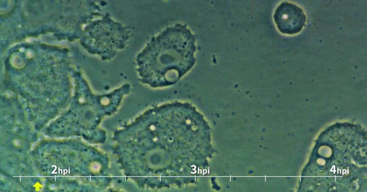
The cells were placed in this chamber under a microscope equipped with a 100x lens and a CCD camera. Through this system, the researchers were able to observe and record the viral infection in real time. At first, Acanthamoeba cells were actively moving across the surface of the culture medium. However, after infection with Mimivirusits movement gradually slowed until it stopped completely. This arrest coincided with the formation of the “virion factory” (VF)a structure within the cytoplasm of the cell that facilitates the production of new viral particles.
Screenshots taken at different times after infection. Source: Journal of Microbiology & Biology Education
The infection videos Mimivirus
The videos present the process of infection of cells of Acanthamoeba for him Mimivirusshowing how the virus invades, replicates and ultimately destroys its host cell. Each video covers a different stage of the viral life cycle, from the first minutes after infection to final cell collapse. The term hpi refers to “hours post infection”. It is used to indicate the time elapsed since the Mimivirus infected the cell. This measurement allows the observation of dynamic changes over time, from the entry of the virus to the release of new viral particles.
Early stages of infection: 1.8 to 3.7 hours post infection (hpi)
In this initial phase, the cells of Acanthamoeba They still show active movement. Entry begins Mimivirusbut changes in the host cell are not yet evident.
Progression of infection: from 3.7 to 6 hours post infection (hpi
The virus begins to replicate inside the cell. It is observed a gradual decrease in cell movementindicating that the infection is progressing and affecting the normal function of the amoeba.
Formation of the “virion factory”: 7.7 to 11.5 hours post infection (hpi)
The creation of the “virion factory“, a specialized compartment where the Mimivirus is assembled. The host cell adopts a more rounded shape, a sign that the virus has taken control.
Final stage and cell rupture: 13.7 to 15.7 hours post infection (hpi)
The life cycle of the virus comes to an end. The Acanthamoeba cell ruptures, releasing numerous viral particles. It is the critical moment where the Mimivirus completes its propagation.
From educational recording to scientific application
The video obtained during the study is an innovative educational tool that has been implemented in biology classrooms at the Tokyo University of Science. According to the authors, seeing the viral replication process helps students visually and dynamically understand the virus life cyclesomething that is often difficult to convey with just static images or diagrams.
The study also offers new insights into the biology of the Mimivirus. The ability to observe the development of the VF and subsequent cell membrane rupture It allowed researchers to document the entire infection cycle, something that had not been possible until now with other smaller viruses. This observation highlights the importance of Mimivirus not only as an object of educational study, but also as a model to better understand the replication mechanisms of complex viruses.
Enlargements of an infected cell, along with diagrams illustrating the proliferation of Mimivirus. Black lines indicate Acanthamoeba cell membranes, while blue circles represent viral particles. Source: Journal of Microbiology & Biology Education
The debate over whether giant viruses are truly living beings
The discovery of Mimivirus and other giant viruses has reopened an old debate in biology: Are viruses really living beings? Traditionally, viruses have been considered non-living entities because they cannot carry out metabolic functions on their own; They depend on a host cell to replicate. However, giant viruses, with their exceptional size and complex genomes, call into question this notion, which for some is simplistic.
He Mimivirus has a genome of double-stranded DNA much larger than that of most viruses, with up to 1.2 million base pairs and more than a thousand coding genes. These genes include some that are typically found in cellular organisms, such as those involved in protein synthesissuggesting that giant viruses might once have had independent biochemical capabilities. According to a study by Claverie and Abergel (2018)the genetic complexity of Mimivirus is comparable to that of many obligate intracellular bacteria, which has led some scientists to propose that giant viruses could represent a fourth domain of life.
On the other hand, other researchers argue that, although giant viruses possess characteristics similar to those of cellular organisms, their absolute dependence on a host to complete their life cycle remains a fundamental criterion that excludes them from the status of living beings. According to Raoult (2004), although giant viruses defy traditional definitions, their inability to replicate outside a host cell reinforces the idea that they are “halfway life forms”, between the living and the nonliving.
This debate is not just academic; has important implications for our understanding of the evolution of life on Earth. Some scientists suggest that giant viruses could be descendants of ancient ways of life that lost the ability to replicate autonomously, becoming obligate parasites. This theory aligns with the concept that giant viruses could have arisen from complex cells that evolved into a parasitic life form, retaining only the genes essential for infection.
References
- Morioka, K., Fujieda, A., Takemura, M. (2024). Visualization of giant Mimivirus in a movie for biology classrooms. Journal of Microbiology and Biology Education, Volume 0, Issue 0.
- Raoult D., Audic S., Robert J., Abergel C., Renesto P., Ogata H., La Scola B., Suzan M., Claverie J.-M. (2004). The 1.2-megabase genome sequence of Mimivirus. Sciencevolume 306, pages 1344–1350.
- Claverie J.-M., Abergel C. (2018). Mimiviridae: an expanding family of highly diverse large dsDNA viruses infecting a wide phylogenetic range of aquatic eukaryotes. Virusesvolume 10, article 506.
- Klose T, Kuznetsov YG, Xiao C, Sun S, McPherson A, Rossmann MG. The three-dimensional structure of Mimivirus. Intervirology. 2010;53(5):268-73. doi: 10.1159/000312911


