THE ESSENTIAL
- Researchers were able to produce the first-ever electron microscopy images of intact coronavirus particles in the olfactory lining.
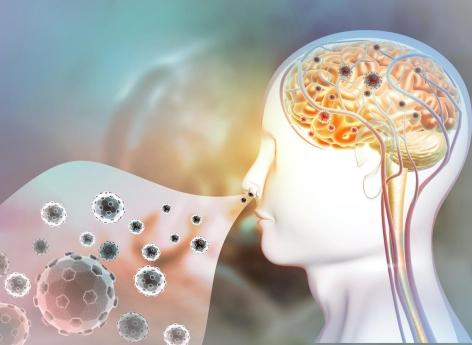
- Once inside the olfactory lining, the virus appears to use neuroanatomical connections, like the olfactory nerve, to reach the brain.
–
Neurological symptoms are reported in more than one in three infected people. It may be loss of smell, taste, headaches, fatigue, dizziness or even nausea. In some patients, the disease can even lead to a stroke or other serious conditions. Using images obtained under an electron microscope, German researchers have managed to trace how the virus enters the central nervous system and subsequently invades the brain. They published their results on November 30 in the journal Nature Neuroscience.
A discovery in severe cases
Observations by researchers at the Charité University Hospital in Berlin revealed that SARS-CoV-2 enters the brain through nerve cells in the olfactory lining. To find out, they analyzed samples taken from the olfactory lining of 33 deceased patients and from four different brain regions. Tissue samples and separate cells were tested for genetic material of SARS-CoV-2 and for a “spike protein” that is found on the surface of the virus. The team provided evidence of the virus in different neuroanatomical structures that connect the eyes, mouth and nose to the brainstem. The olfactory mucosa is the one that showed the highest viral load. Researchers were able to produce the first-ever electron microscopy images of intact coronavirus particles in the olfactory lining.
“These data support the idea that SARS-CoV-2 is able to use the olfactory lining as a gateway to the brain.”, Concluded Professor Frank Heppner who led the team of multidisciplinary researchers. This is also supported by the close anatomical proximity of the mucous cells, blood vessels and nerve cells in the area. “Once inside the olfactory lining, the virus appears to use neuroanatomical connections, like the olfactory nerve, to reach the brain, added the neuropathologist. It is important to point out, however, that the Covid-19 patients involved in this study had what would be defined as serious illness, belonging to that small group of patients in whom the disease is fatal. It is therefore not necessarily possible, therefore, to transfer the results of our study to cases of mild or moderate disease..”
Respiratory function also affected
How the virus travels through nerve cells remains to be discovered. “Our data suggest that the virus travels from one nerve cell to another to reach the brain, says Dr Helena Radbruch who participated in the study. It is likely, however, that the virus is also transported via blood vessels as evidence of the virus has also been found in the walls of blood vessels in the brain..”
Researchers have also studied how the immune system responds to infection. In addition to finding evidence of activated immune cells in the brain and in the olfactory lining, they detected the immune signatures of these cells in brain fluid. In some of the cases studied, the researchers also found tissue damage caused by a stroke as a result of thromboembolism, which is the obstruction of a blood vessel by a blood clot.
“In our view, the presence of SARS-CoV-2 in the nerve cells of the olfactory mucosa provides a good explanation for the neurological symptoms encountered in Covid-19 patients, such as a loss of smell or taste., concluded Prof. Heppner. We also found SARS-CoV-2 in areas of the brain that control vital functions, such as breathing. It cannot be ruled out that, in patients with severe Covid-19, the presence of the virus in these areas of the brain will have an exacerbating impact on respiratory function, adding to respiratory problems due to SARS lung infection. -CoV-2. Similar problems could arise in relation to cardiovascular function.”
<!–
Ce sujet vous intéresse ? Venez en discuter sur notre forum !
–


